When Is the Right Time to Remove a Skin Tag or Mole?
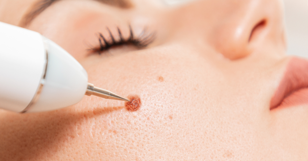
Deciding when to remove a skin tag or mole can be tricky, especially if you’re unsure about the signs to watch for. If you notice changes in size, colour, or shape, it might be time to reflect on removal. These changes would indicate potential health concerns. On the other hand, if a lesion causes irritation, discomfort, or affects your self-esteem, seeking removal could improve your quality of life. Regular self-examinations and consultations with a dermatologist are essential. But how do you know which professional removal methods are right for you? Let’s explore your options. Key Takeaways – Removal is advised if the mole or skin tag shows rapid growth or significant size changes. – Consider removal if the lesion changes colour, especially if it turns dark or multi-colored. – Seek medical evaluation if the skin tag or mole bleeds or oozes without apparent cause. – Persistent itching, pain, or irritation from the lesion indicates it may need to be removed. – A mole with irregular shape or asymmetrical borders should be examined and potentially removed. Cosmetic Reasons Considering the cosmetic reasons for removing a skin tag or mole, it’s essential to understand the impact on one’s self-esteem and appearance. Skin aesthetics play a significant role in how individuals perceive themselves and are perceived by others. If a skin tag or mole is located in a prominent area such as the face or neck, it can lead to self-consciousness, affecting social interactions and confidence levels. Cosmetic procedures aimed at removing these skin imperfections are generally safe and effective. Techniques such as cryotherapy, laser removal, and surgical excision are commonly used. These methods not only improve skin aesthetics but also provide lasting results with minimal scarring. Clinical studies have shown that individuals who undergo these procedures often experience a boost in self-esteem and overall satisfaction with their appearance. It’s important to consult with a dermatologist to assess the best method for removal based on the size, location, and type of skin tag or mole. This guarantees that the procedure is tailored to your specific needs, maximising both cosmetic and psychological benefits. By addressing these concerns through professional cosmetic procedures, you can enhance your skin aesthetics and overall quality of life. Irritation and Discomfort While cosmetic reasons play an important role in the decision to remove a skin tag or mole, irritation and discomfort often necessitate medical intervention. Friction from clothing, jewellery, or even daily activities can exacerbate these skin lesions, leading to inflammation and potential secondary infections. When you experience persistent irritation, it’s essential to consult a healthcare provider to discuss removal options. Self care practices are vital in managing irritated skin tags or moles. Applying a gentle, non-irritating moisturiser can help reduce friction and soothe the skin. Additionally, wearing loose-fitting clothing can minimise contact and irritation. However, these measures might only offer temporary relief. Chronic discomfort or irritation can greatly impact your overall skin health. Prolonged inflammation not only causes pain but also increases the risk of infection. If you notice that a skin tag or mole consistently causes discomfort despite your self care efforts, it’s time to seek professional advice. A dermatologist can provide evidence-based treatments such as cryotherapy, excision, or laser removal, ensuring the best outcome for your skin health. Don’t ignore signs of irritation. Addressing these issues promptly can prevent further complications and improve your quality of life. Changes in Size One of the most concerning changes in a skin tag or mole is an increase in size. Monitoring changes in size progression is essential because it can be an indicator of underlying health issues, including malignancy. Clinically, a rapid or continuous increase in size warrants a thorough evaluation by a healthcare professional. You should regularly examine your skin tags and moles, noting any size progression. Use a ruler or take pictures monthly to document any changes. If you observe considerable growth, schedule an appointment with a dermatologist. They may perform a biopsy to rule out conditions like melanoma, which presents with asymmetric borders and rapid growth. Evidence suggests that size progression, particularly when accompanied by other symptoms like bleeding or itching, is a red flag. A study published in the Journal of the American Academy of Dermatology highlights that early detection and removal of suspicious moles can greatly improve outcomes. Don’t ignore changes in size, even if they seem minor. Early intervention is key. By actively monitoring changes and consulting with a healthcare provider, you can guarantee timely and appropriate management, safeguarding your health. Colour Alterations Over time, alterations in the colour of a skin tag or mole can signal important health concerns. You should pay close attention to any changes in hue, as these could indicate underlying issues affecting your skin health. For example, a mole that darkens or develops multiple colours, such as shades of brown, black, red, or blue, might be a red flag. These colour meanings often point to melanocytic activity, which could be benign or malignant. Don’t ignore a mole that suddenly becomes lighter or loses pigment. This depigmentation might suggest regression, a phenomenon sometimes seen in malignant melanomas. Clinicians use the “ABCDE” rule to assess moles: Asymmetry, Border irregularity, Color variation, Diameter over 6mm, and Evolution. Colour variation, in particular, is a vital indicator that warrants professional evaluation. It’s essential to monitor your skin regularly and consult a dermatologist if you notice any colour changes. Early detection and intervention can greatly improve outcomes. Shape Modifications When a skin tag or mole starts shifting its shape, it’s time to pay attention. Shape variations can be a critical indicator of underlying health issues. While some changes might be benign, others could signal more serious conditions such as melanoma. Here are some key points to take into account: Asymmetry: If one half of the mole or skin tag doesn’t match the other half,
How Effective Is Mole Removal in Reducing Skin Cancer Risk?
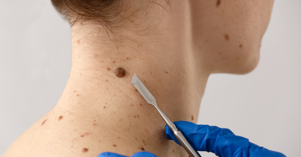
When considering the effectiveness of mole removal in reducing skin cancer risk, you should first understand the types of moles that pose a threat. Spotting suspicious moles early and opting for procedures like excisional surgery can indeed lower potential risks. However, you might wonder if these methods are foolproof and what role accurate diagnosis plays in the overall strategy. Are there more preventive measures you could take beyond just removing moles? Key Takeaways – Mole removal is highly effective when suspicious moles are accurately diagnosed and excised early. – Excisional and Mohs surgeries offer precise removal, minimising the risk of cancerous cells remaining. – Regular follow-up dermatologic check-ups post-removal are crucial for monitoring and reducing skin cancer risk. – ABCDE criteria and biopsy confirmation prior to removal ensure the identification of high-risk moles. – Early intervention through mole removal significantly reduces the likelihood of moles progressing to skin cancer. Understanding Moles Although moles are a common dermatologic occurrence, understanding their characteristics is essential for effective monitoring and management. Moles, or nevi, typically present as small, pigmented lesions on the skin. Their characteristics include symmetry, uniform colour, distinct borders, and a diameter usually less than 6mm. However, any deviation from these traits warrants closer attention. When it comes to mole monitoring, you should perform regular self-examinations using the ABCDE criteria: Asymmetry, Border irregularity, Color variation, Diameter over 6mm, and Evolving shape or size. This evidence-based approach helps you identify atypical moles that might require professional evaluation. Dermatologists often recommend photographic documentation for mole monitoring, especially if you have numerous moles or a history of atypical nevi. High-resolution images can serve as a baseline, allowing you and your healthcare provider to track any changes over time. If you notice any new symptoms such as itching, bleeding, or rapid growth, seek medical advice immediately. Types of Skin Cancer Spotting atypical moles early is important, as they can sometimes be precursors to more severe skin conditions. Among the various types of skin cancer, understanding the differences is essential for effective prevention and treatment. The most dangerous form is melanoma. There are several melanoma types, including superficial spreading melanoma, which is the most common and often appears as a flat or slightly raised discoloured patch with irregular borders. Another type, nodular melanoma, presents as a rapidly growing bump that can be black, blue, or red. Basal cell carcinoma (BCC) is the most common skin cancer but also the least aggressive. BCCs often manifest as pearly or waxy bumps, usually on sun-exposed areas like the face or neck. They rarely metastasize but can cause significant local damage if left untreated. Squamous cell carcinoma (SCC) is another prevalent skin cancer, often arising from prolonged sun exposure. SCCs appear as rough, scaly patches or nodules that can become crusted or bleed. The Procedure of Mole Removal Mole removal, an important dermatological procedure, involves excising suspicious or bothersome moles to prevent potential malignancies and improve skin health. To begin, your dermatologist will evaluate the mole using dermatoscopy to determine the best approach. Several surgical techniques are employed based on the mole’s characteristics and location. For superficial moles, shave excision is a common method. Here, the dermatologist uses a scalpel to shave the mole flush with your skin. For deeper or potentially malignant moles, an elliptical excision might be used. This involves cutting around the mole and removing a margin of healthy tissue to guarantee complete excision. Occasionally, a punch biopsy, which uses a circular blade to remove a cylindrical core of skin, is utilised for smaller moles. Post-procedure, the healing process is vital. Your dermatologist will likely provide specific wound care instructions to minimise infection risk and promote ideal healing. Typically, the area will heal within one to two weeks, though it may take longer depending on the mole’s size and depth. Follow-up appointments guarantee proper healing and evaluate if further treatment is necessary. Adhering to these guidelines will help maintain skin integrity and reduce complications. Effectiveness of Mole Removal The effectiveness of mole removal hinges on several key factors, including the accuracy of the initial diagnosis and the chosen surgical technique. First, identifying mole characteristics is vital. Dermatologists often use the ABCDE criteria—Asymmetry, Border irregularity, Color variation, Diameter over 6mm, and Evolving shape—to assess malignancy risk. A biopsy, if warranted, confirms whether a mole is benign or malignant. This step guarantees that only moles with suspicious characteristics are targeted for removal, minimising unnecessary procedures. Once a mole is identified for removal, the surgical technique plays a notable role. Methods like excisional surgery or Mohs surgery offer high precision and efficacy. Excisional surgery involves removing the mole along with a margin of healthy skin, which is particularly effective for potentially malignant moles. Mohs surgery, often reserved for confirmed skin cancers, allows for layer-by-layer removal and immediate microscopic examination, guaranteeing complete excision while sparing healthy tissue. Post-removal skin monitoring is essential for long-term effectiveness. Regular dermatologic check-ups help identify new moles or changes in existing ones, providing an ongoing defence against skin cancer. Alternatives to Mole Removal While surgical removal of moles remains a highly effective method, various non-surgical alternatives also warrant consideration, particularly for benign moles or patients seeking less invasive options. Here are some evidence-based alternatives you might explore: Topical Treatments: Prescription creams containing imiquimod or fluorouracil can be applied to the mole under medical supervision. These agents work by stimulating the immune system to target abnormal cells. Cryotherapy: This method involves freezing the mole with liquid nitrogen, causing the abnormal tissue to die and eventually fall off. It’s less invasive than surgical removal and can be effective for superficial moles. Laser Therapy: Using concentrated light, laser therapy targets pigmented cells in the mole. It’s particularly beneficial for flat moles and those in cosmetically sensitive areas. Home Remedies and Monitoring: While home
What Should You Expect During and After Mole Removal?
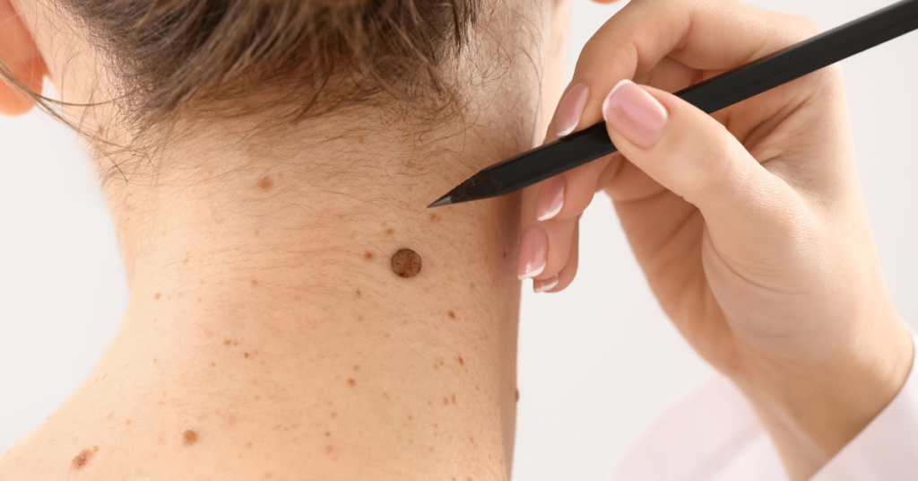
Navigating mole removal is like starting on a journey through a well-charted map. You’ll begin with an initial consultation, where a dermatologist assesses the mole and discusses your options. The procedure itself, whether a surgical excision or laser therapy, involves local anaesthesia to guarantee comfort. Afterward, you’ll need to keep the wound clean and watch for any signs of infection. Healing times can vary, and follow-up appointments are essential to monitor any changes in your skin. Curious about how to manage pain or what long-term care involves? Let’s explore the steps in detail. Key Takeaways – Dermatologists assess mole type and medical history before choosing a removal technique. – Local anaesthetic is administered to numb the area during the procedure. – Post-procedure care includes keeping the wound clean, using prescribed ointments, and monitoring for infection. – Healing time varies but typically takes 1-3 weeks, depending on the removal method. – Regular follow-up visits and skin checks are crucial for monitoring and preventing future skin issues. Initial Consultation During your initial consultation, you’ll meet with a dermatologist to discuss your mole removal options. This meeting is vital for evaluating the types of moles you have and determining the best treatment plan. The dermatologist will begin by conducting a thorough skin evaluation. They’ll examine the size, shape, colour, and texture of your moles to identify whether they’re benign or potentially malignant. Understanding the different mole types is essential in this evaluation process. Common mole types include junctional moles, which appear flat and dark, compound moles, which are slightly raised and lighter in colour, and intradermal moles, which are raised and flesh-coloured. Each type requires a different approach for removal, so accurate identification is key. The dermatologist will also inquire about your medical history, including any family history of skin cancer, to assess your overall risk. This helps in creating a personalised treatment plan tailored to your needs. They’ll explain the various removal techniques, such as surgical excision, laser therapy, or cryotherapy, and discuss the benefits and potential risks associated with each method. Pre-Procedure Preparations Before undergoing mole removal, there are several key preparations you’ll need to make to guarantee a smooth procedure. First, your healthcare provider will conduct a thorough mole assessment. This helps them understand the characteristics of your mole and determine the best removal method for your specific skin type. Different skin types react differently to procedures, so this step is essential. Here’s a checklist to help you prepare: Consultation Review: Revisit the details discussed during your initial consultation to confirm you understand the procedure and aftercare. Medication Disclosure: Inform your doctor about any medications or supplements you’re taking as they may affect the procedure or healing process. Skin Preparation: Follow any specific skin preparation instructions your doctor provides, such as avoiding certain skincare products or sun exposure. Pre-Procedure Instructions: Adhere to any fasting or hydration guidelines given by your healthcare provider. Allergy Check: Confirm you’re not allergic to any materials that will be used during the procedure, such as anaesthetics or antiseptics. The Mole Removal Process The mole removal process typically begins with the administration of a local anaesthetic to numb the area, guaranteeing you won’t feel any discomfort during the procedure. Once the area is numb, your healthcare provider will determine the most appropriate removal technique based on the type of mole you have. There are various mole types, including common moles, atypical moles, and congenital moles. Each type may require a different approach. For instance, a simple shave excision is often used for small, raised moles. In this technique, the mole is carefully shaved off at the skin’s surface. If your mole is flat or suspected to be atypical, a punch biopsy might be employed. This method involves using a small, round blade to remove a core of skin, including the mole and some surrounding tissue. For larger or deeper moles, a full-thickness excision might be necessary. This involves cutting out the mole along with a margin of healthy skin, followed by suturing the wound. After the mole is removed, it’s typically sent to a lab for histopathological examination to rule out malignancy. This step is essential, especially for atypical moles, to safeguard your long-term health. Immediate Post-Procedure Care Your healthcare provider’s immediate post-procedure care instructions are essential to ascertain proper healing and prevent complications. Following these guidelines meticulously will help you manage pain and guarantee peak wound care. Here are the key steps you should take: – Keep the wound clean and dry: Gently clean the area with mild soap and water, then pat it dry. Avoid soaking the wound or exposing it to excessive moisture. – Apply prescribed ointments: Use any topical antibiotics or healing ointments as directed to prevent infection and promote healing. – Change bandages regularly: Replace the dressing daily or more often if it becomes wet or dirty. This helps keep the wound clean and reduces the risk of infection. – Monitor for signs of infection: Watch for redness, swelling, increased pain, or discharge. If you notice any of these symptoms, contact your healthcare provider immediately. – Manage pain effectively: Use over-the-counter pain relievers like acetaminophen or ibuprofen as recommended. Avoid aspirin, as it can increase bleeding. Healing and Recovery Timeline Typically, the healing and recovery timeline for mole removal can vary depending on the method used and individual factors such as your overall health and skin type. For instance, if you’d a surgical excision, it may take about two to three weeks for the wound to heal. On the other hand, laser removal often results in a faster recovery, sometimes within a week. Key healing factors include your body’s natural healing ability and how well you follow post-procedure care instructions. Keeping the area clean and dry, applying prescribed ointments, and avoiding direct sunlight
What Are the Key Differences Between Skin Tag Removal and Mole Removal?
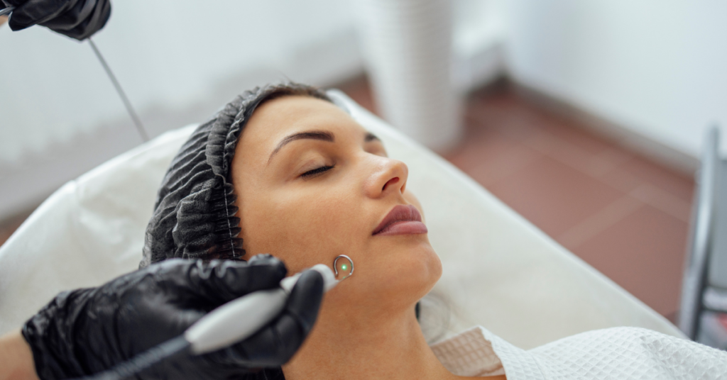
When you’re considering skin tag removal versus mole removal, you’ll notice distinct differences in their characteristics and the techniques used. Skin tags are typically small, flesh-coloured growths caused by friction, often removed with cryotherapy or electrosurgery, leading to minimal scarring and a quick recovery. On the other hand, moles are pigmented lesions that may necessitate excisional surgery or laser treatment, involving deeper tissue and more extensive aftercare. Understanding these differences can help you make informed decisions about which procedure might be best for you. So, what else should you know before deciding on a treatment path? Key Takeaways – Skin tags are typically removed due to friction and discomfort, while moles are often monitored for potential malignancy. – Skin tag removal methods include cryotherapy and electrosurgery; mole removal methods include laser techniques and excisional surgery. – Skin tag removal generally involves minimal pain and quick recovery; mole removal can be more painful and have a longer healing time. – Professional removal of moles is crucial to accurately assess for malignancy, whereas skin tags are usually benign. – Mole removal often results in more noticeable scarring compared to the usually minimal scarring from skin tag removal. Identification and Characteristics When identifying skin tags and moles, it’s vital to understand their distinct characteristics to guarantee accurate treatment. Skin tag characteristics typically include small, soft, flesh-coloured or slightly darker growths that hang off the skin by a thin stalk, known as a peduncle. They’re generally painless and can appear on areas prone to friction, such as the neck, armpits, and groyne. Skin tags are benign and don’t usually transform into malignant lesions. In contrast, mole characteristics can vary considerably. Moles, or nevi, are pigmented lesions that can be flat or raised, and their colour ranges from tan to dark brown or black. They’re often round or oval with a smooth border. Moles can appear anywhere on the body and may change over time. While most moles are benign, some can develop into melanoma, a type of skin cancer. As a result, it’s important to monitor moles for changes in size, shape, colour, or symmetry, as these could indicate malignancy. Causes and Risk Factors Understanding the causes and risk factors for skin tags and moles is essential for appropriate skin health management. Skin tags, known medically as acrochordons, often arise due to friction where skin rubs against skin or clothing. Obesity and type 2 diabetes are significant risk factors. Genetic factors also play a role; if your family members have skin tags, you’re more likely to develop them too. Moles, or nevi, are primarily influenced by genetic factors. If you have a family history of moles, you might see them more frequently on your skin. Ultraviolet (UV) radiation from sun exposure is a critical environmental trigger for mole development and changes. This environmental factor can activate melanocyte cells, increasing the likelihood of moles forming or existing ones changing in appearance. Both skin tags and moles can be exacerbated by hormonal changes, particularly during pregnancy. While moles and skin tags are generally benign, understanding these causes and risk factors can help you monitor your skin more effectively. Keep an eye on any new growths or changes to existing skin features and consult a dermatologist for personalised advice based on your genetic predispositions and environmental exposures. Common Removal Techniques Removing skin tags and moles involves a variety of clinical techniques tailored to the specific nature of each growth. When you’re dealing with skin tags, cryotherapy methods are commonly employed. This entails applying liquid nitrogen to freeze the tag, causing it to fall off naturally within a few days. Cryotherapy is minimally invasive and generally well-tolerated. For mole removal, laser techniques are often preferred. A concentrated beam of light is used to target the pigmented cells in the mole, effectively vaporising them. Laser techniques offer precision and minimal scarring, making them suitable for moles in sensitive areas such as the face. However, you’ll need to verify the mole is benign before opting for this method. Electrosurgery is another option for both skin tags and moles. This technique uses an electric current to burn off the growth. It’s quick and efficient but requires local anaesthesia. Additionally, excisional surgery, which involves cutting out the mole or tag, is considered when larger or suspicious growths are present. Each technique has its specific indications and benefits, so consult a dermatologist to determine the most appropriate method for your situation. Understanding these options helps you make an informed decision about your skin health. Professional vs. DIY Methods In the domain of skin growth management, opting for professional intervention over DIY methods offers significant advantages. Professional techniques are executed by dermatologists who use sterile equipment and advanced technology, ensuring a higher success rate and minimal scarring. They can accurately distinguish between benign and malignant growths, a critical step in preventing potential health risks. Conversely, DIY methods often lack the precision and safety measures necessary for effective removal, leading to complications like infection or incomplete excision. When you choose professional techniques for skin tag or mole removal, you’re benefiting from clinical expertise and rigorous sanitation standards. Cryotherapy, laser removal, and surgical excision are some methods that provide reliable outcomes with reduced recurrence rates. These procedures are backed by evidence-based practices, ensuring both efficacy and patient safety. On the flip side, DIY methods, such as over-the-counter creams or at-home kits, pose significant risks. Without professional assessment, you might misidentify a mole as benign when it’s actually malignant, delaying essential medical care. Additionally, improper use of DIY methods can result in suboptimal healing, increased scarring, and potential secondary infections. Consequently, while DIY methods might seem convenient, the benefits of professional techniques far outweigh the risks. Pain and Discomfort Levels Have you ever wondered how much pain and discomfort you’ll experience during skin tag or
Is Mole Removal Safe and Effective With Astrum Medical?
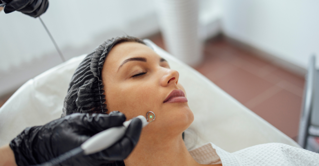
You might not know that Astrum Medical uses some of the most advanced diagnostic tools in the industry for mole removal. Their specialised training and innovative techniques aim to maximise both safety and effectiveness. You’ll find that they prioritise patient comfort through thorough consultations, strict sterilisation measures, and the use of local anaesthesia. But what about their success rates, patient outcomes, and potential risks? To understand how Astrum Medical stands out from other providers and whether their approach is right for you, there’s more you should consider. Key Takeaways – Astrum Medical uses advanced diagnostic tools and minimally invasive techniques for precise and effective mole removal. – Strict safety protocols, including sterilisation and local anaesthesia, ensure a safe procedure at Astrum Medical. – Techniques like laser ablation and cryotherapy promote faster recovery and minimise discomfort. – Over 95% of patients report favourable cosmetic results and minimal scarring post-procedure. – Board-certified dermatologists at Astrum Medical bring extensive experience, enhancing the safety and effectiveness of mole removal. Understanding Mole Removal Understanding mole removal begins with recognising the different types of moles and the medical criteria used to evaluate whether they should be excised. Moles, or nevi, can be congenital or acquired, and they vary considerably in size, shape, and colour. Dermatologists typically assess moles using the ABCDE criteria—Asymmetry, Border irregularity, Color variation, Diameter larger than 6mm, and Evolving characteristics. These factors help determine if a mole is benign or potentially malignant, which informs the decision to remove it. When considering mole removal, you’ll need to weigh both medical and cosmetic concerns. Medically, excision may be necessary to prevent malignancy. Cosmetically, moles in visible areas can affect self-esteem and appearance. The healing process is a critical aspect of mole removal. Post-excision, the wound undergoes a sequence of healing phases: hemostasis, inflammation, proliferation, and maturation. Proper wound care is essential to minimise scarring, which is often a primary cosmetic concern for patients. Evidence-based practices dictate the use of sterile techniques and potential application of topical agents to enhance the healing process. Understanding these facets guarantees you make informed decisions about mole removal, balancing health risks and cosmetic outcomes effectively. Astrum Medical’s Approach Astrum Medical uses a cutting-edge, patient-centred methodology for mole removal, integrating advanced diagnostic tools and minimally invasive techniques to secure best results. By leveraging innovative technology like high-resolution dermoscopy and digital mole mapping, they can accurately assess and monitor moles with exceptional precision. This ensures that only moles requiring removal are targeted, minimising unnecessary procedures and optimising patient outcomes. Their team undergoes specialised training in the latest dermatological advancements and surgical techniques, securing they’re well-equipped to handle complex cases. This rigorous training includes proficiency in laser ablation, radiofrequency excision, and cryotherapy, which are less invasive and promote faster recovery compared to traditional surgical methods. Moreover, Astrum Medical’s clinicians employ state-of-the-art equipment that enhances the efficacy and safety of these procedures. Evidence-based practice is a cornerstone of their approach, securing that each treatment plan is tailored to the specific needs of the patient. By continuously updating their protocols based on the latest research and clinical guidelines, Astrum Medical maintains its commitment to providing high-quality care. This combination of innovative technology and specialised training secures that you receive the most effective and safest mole removal treatment available. Safety Protocols Safety measures at Astrum Medical are carefully crafted to protect patients from potential complications, guaranteeing a safe and efficient mole removal experience. The team prioritises strong safety precautions, starting with a thorough initial consultation. During this phase, your medical history, allergies, and any medications you’re taking are meticulously reviewed. This pre-procedure assessment helps identify any risk factors that could complicate the mole removal process. On the day of the procedure, the medical staff follows strict sterilisation protocols. All instruments are sterilised using cutting-edge autoclaves, and the surgical area is prepared to minimise any risk of infection. Local anaesthesia is administered to ensure your comfort, and the mole removal is conducted under aseptic conditions, significantly decreasing the likelihood of post-procedure complications. Post-procedure care is equally crucial. You’ll receive detailed instructions on wound care, including how to keep the area clean and protected. Follow-up appointments are scheduled to monitor the healing process and address any concerns promptly. This thorough approach to post-procedure care guarantees optimal healing and minimises the risk of infection or scarring. Techniques Used Frequently, mole removal at Astrum Medical employs advanced techniques such as laser therapy, cryotherapy, and surgical excision to guarantee precision and effectiveness. Each method offers unique benefits tailored to the specific characteristics of the mole and the patient’s skin type. Laser Treatment: This technique uses concentrated light energy to break down pigmented cells in the mole. It’s highly precise, minimising damage to surrounding tissue and reducing the risk of scarring. Laser treatment is particularly effective for removing flat or slightly raised moles. Cryotherapy: This method involves freezing the mole with liquid nitrogen, causing the cells to die and fall off. Cryotherapy is ideal for superficial moles and offers a quick, non-invasive solution with minimal discomfort. Surgical Excision: In cases where the mole is large, irregular, or potentially malignant, surgical excision is the preferred technique. This involves cutting out the mole with a scalpel and stitching the area closed. Surgical excision ensures complete removal and allows for histopathological examination. Shave Excision: For raised moles that aren’t deeply rooted, shave excision involves shaving off the mole with a blade. This method is less invasive than full surgical excision and usually requires no stitches. These evidence-based techniques guarantee that mole removal at Astrum Medical is both safe and effective. Expert Opinions Many dermatologists and medical experts agree that the advanced techniques utilised by Astrum Medical guarantee high efficacy and patient satisfaction in mole removal procedures. Leveraging cutting-edge technology and specialised medical expertise, Astrum Medical ensures that each procedure is meticulously executed to maximise outcomes and minimise complications.
How Does Mole Removal Work at Astrum Medical?
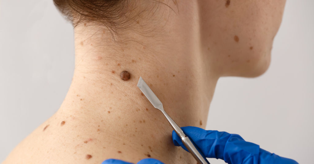
It’s interesting that you’re thinking about mole removal, as we at Astrum Medical have our own way of doing it. You’ll start with an in-depth consultation where a dermatologist evaluates your mole and discusses the best removal options. Whether it’s excision, laser therapy, or cryotherapy, each method has its pros and cons. The focus is always on safety and achieving the best cosmetic results. But what happens after the procedure? There’s a tailored recovery plan you’ll need to follow meticulously. Let’s explore the steps involved and what you can expect during the healing process. Key Takeaways – Dermatologists assess moles and discuss removal options like excision, laser therapy, and cryotherapy. – Mole mapping and digital imaging track changes over time for effective monitoring. – Removal techniques prioritise safety, scar prevention, and cosmetic results using methods like surgical excision and laser treatment. – Local anaesthetics are used for pain management during procedures. – Post-treatment care and follow-up appointments ensure proper healing and long-term monitoring. Initial Consultation During your initial consultation at Astrum Medical, the dermatologist will assess your mole and discuss potential removal options tailored to your specific needs. They’ll consider factors like the mole’s size, location, and appearance. This thorough approach ensures that you receive the most effective treatment options available. Your dermatologist will explain each method, including excision, laser therapy, and cryotherapy, highlighting the benefits and risks of each. Addressing patient concerns is a top priority. You’ll have the opportunity to ask questions about the procedure, recovery time, and potential scarring. The dermatologist will provide detailed answers to help you make an informed decision. They understand that each patient has unique concerns and will take the time to discuss any specific issues you may have, such as pain management or cosmetic outcomes. Skin Examination The dermatologist will meticulously inspect your skin to evaluate the mole’s characteristics and determine the most appropriate elimination method. During your dermatologist consultation, the specialist will use a dermatoscope to examine the mole’s size, shape, colour, and border. This thorough examination helps identify any unusual features that may indicate malignancy. As part of melanoma screening, the dermatologist will assess the mole for asymmetry, irregular borders, multiple colours, diameter greater than 6mm, and any evolution over time—commonly known as the ABCDE criteria. These factors are vital in distinguishing benign moles from potential melanomas. If any signs raise concern, the dermatologist may recommend a biopsy to obtain a definitive diagnosis before proceeding with removal. Your medical history and any family history of skin cancer will also be discussed to provide a comprehensive evaluation. This information helps in understanding your risk factors and tailoring the approach accordingly. Mole Mapping Through mole mapping, a dermatologist creates a detailed record of your moles to monitor for any changes over time, ensuring early detection of potential skin issues. This process involves a thorough initial examination where each mole’s location, size, colour, and shape are documented. Digital imaging technology plays a pivotal role in this procedure, allowing your dermatologist to capture high-resolution images of your skin. These images are then stored in a secure database for future comparison. During follow-up appointments, your dermatologist will refer to these baseline images to assess any changes in your moles. This methodical approach to monitoring progress is vital for identifying early signs of melanoma or other skin abnormalities. By comparing current images with previous ones, even subtle changes can be detected promptly. Mole mapping is particularly beneficial for individuals with a high number of moles or those with a personal or family history of skin cancer. It provides a detailed overview of your skin’s condition, helping your dermatologist make informed decisions about your care. Regular mole mapping appointments are a proactive step in maintaining your skin health and catching potential issues before they become serious. Removal Techniques At Astrum Medical, mole removal techniques are tailored to suit each patient’s unique needs, guaranteeing both safety and effectiveness. You’ll find that our approach prioritises scar prevention and excellent cosmetic results. We offer several methods, including laser removal, cryotherapy, and shave excision, each chosen based on the mole’s characteristics and your personal preferences. Laser removal employs focused light beams to target pigmented cells, minimising damage to surrounding tissue and reducing scarring. Cryotherapy involves freezing the mole with liquid nitrogen, causing it to fall off naturally. Shave excision, on the other hand, involves carefully shaving the mole off the skin’s surface, ideal for raised moles. Pain management is crucial to our process. We use local anaesthetics to ensure you remain comfortable during the procedure. Post-treatment care is equally important; you’ll receive detailed instructions to help your skin heal properly and minimise any potential scarring. This includes keeping the area clean, applying prescribed ointments, and avoiding sun exposure. Cosmetic concerns are addressed during your consultation, where we discuss the best technique for achieving your desired outcome. At Astrum Medical, we aim to provide you with effective results while prioritising your comfort and care. Surgical Excision For moles that require a more invasive approach, surgical excision offers a precise and effective solution. At Astrum Medical, this procedure involves removing the mole along with a margin of surrounding tissue to ensure complete excision. Your physician will use a local anaesthetic to numb the area, making the process virtually painless. Scar prevention is a primary focus during surgical excision. Our skilled surgeons employ advanced techniques to minimise scarring, such as precise incision lines and careful suturing. After the mole is removed, the incision is closed with fine stitches, which are usually removed within one to two weeks. Post-operative care is essential for best healing and scar prevention. You’ll receive detailed instructions on how to care for the wound, including keeping the area clean and dry, applying prescribed ointments, and avoiding strenuous activities that may strain the incision. Follow-up appointments are scheduled to monitor the healing process and address


